A study by researchers from KTH explores Lupinus angustifolius (a type of lupin) as a sustainable source of lignocellulose materials. In order to reduce reliance on forest wood and utilise agricultural residues to produce microfibrillated cellulose (MFC), the team analysed the anatomical structure and chemical composition of the plant, classifying parts into stems, roots, seed pods, and seeds and developed a mild extraction process using alkaline treatment to create lignin-containing MFC (L-MFC) from non-edible residues.
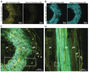
The researchers employed several methods to track changes during extraction: they used scanning electron microscopy (SEM) and Fourier-transform infrared spectroscopy (FTIR) to examine the plant's morphology and chemical transformations. Using Carbotrace 680 allowed them to map cellulose distribution in native tissues and throughout the extraction. By soaking tissue sections in Carbotrace and analysing them under a confocal microscope, they visualised the spatial relationships between cellulose and lignin in real-time; this provided critical insights into the distribution of biomacromolecules across various tissues.
The study concluded that Lupinus angustifolius is a viable alternative source for lignocellulose materials. The mild extraction process successfully separated cellulose while preserving lignin, which enhanced the strength and thermal properties of the final product; Carbotrace enabled precise, non-invasive visualization of cellulose, guiding efficient lignocellulose recovery throughout the process. This research highlights the potential of agricultural residues for producing bio-based materials, offering a sustainable alternative to forest-derived resources.
Image: Confocal fluorescence microscopy images of Lupinus angustifolius tissues stained with Carbotrace 680 to highlight the distribution of lignin and cellulose. Panels (A) and (B) display cross-sections with and without Carbotrace 680. In (A), excitation at 561 nm (emission 570-631 nm) highlights cellulose in yellow fluorescence, while the control shows no signal, confirming the stain’s specificity. Panel (B) shows lignin in blue (excitation 405 nm, emission 406-505 nm), for which Carbotrace does not stain, showing there is autofluorescence with and without the Carbotrace. Panels (C) and (H) illustrate cross-sections and longitudinal sections of the stem, showing the distribution of lignin and cellulose in tissues like xylem (xy), phloem (ph), and pith (pi). Insets in (C) and (H) provide detailed views of these regions. Image from Figure 2A,B,C and H by Schmidt et al 2024, Advanced Sustainable Systems, 8, 2300632 (CC BY 4.0).