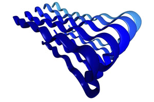
Copper’s role in prion protein misbehaviour
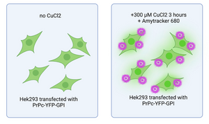
Prion diseases are fatal neurodegenerative disorders caused by the misfolding and aggregation of prion proteins (PrP). Although copper (Cu2+) imbalance has long been suspected to play a role in these conditions, a recent study by Juliani and colleagues at the Federal University of Rio de Janeiro sheds new light on this issue. Their researchreveals that Cu2+ promotes the formation of liquid-like PrP condensates that naturally buffer excess copper and help prevent toxicity. However, under oxidative stress, these condensates transition from a liquid to solid state, forming amyloid-like aggregates that may contribute to disease progression. These findings suggest a critical link between Cu2+ dysregulation, oxidative stress, and prion pathology.
Detecting and characterizing prion aggregates is key to understanding their impact on neurodegeneration. In this study, the researchers used Amytracker 680 to visualize amyloid-like PrP aggregates in Cu2+-treated cells, confirming that prolonged exposure to Cu2+ leads to PrP aggregation - a process that mirrors the pathological changes seen in prion diseases.This work not only highlights the potential of Amytracker as a tool for tracking protein aggregation, but also supports further research into neurodegenerative disorders and potential therapeutic strategies.
Image: Figure illustrating live-cell imaging of HEK293 cells expressing PrPC-YFP-GPI. Cells treated with 300 μM CuCl2 for 3 hours (right panel) show AmyTracker680-positive aggregates on the cell surface. Image created using BioRender.
Read More:
How functional amyloids help regulate stress responses
Protein aggregation is traditionally seen as a pathological process, hallmark of neurodegenerative diseases and systemic amyloidoses. However, emerging evidence reveals that amyloid aggregation is not merely the consequence of protein misfolding; rather, cells harness reversible amyloid aggregation to adapt to environmental stress. Understanding how functional amyloids form and disassemble could provide critical insights into the treatment of pathological aggregation.
The team led by Timothy Audas at Simon Fraser University, has recently published its work on Amyloid bodies (A-bodies) - nuclear amyloid-like inclusions that sequester proteins in response to environmental stressors. Similar to other stress-related inclusion (e.g. cytoplasmic stress granules) the composition of A-bodies is stress-specific. This suggests that cells dynamically regulate the recruitment of proteins into A-bodies based on environmental conditions. Notably, several disease-associated proteins, such as β-amyloid peptides, are recruited and aggregate within A-bodies, indicating a potential link between pathological amyloidogenesis and dysregulation of A-body formation.
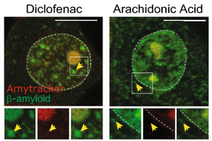
The study highlights how small molecules that reduce the aggregation of β-amyloid in vitro had no effect on the recruitment of pathological fragments A-bodies. On the other hand, diclofenac, a nonsteroidal anti-inflammatory drug, despite having no effect in traditional in vitro assays, significantly reduced the amount of β-amyloid aggregates formed in response to stress. Importantly, the drug does not impair the broader formation of A-bodies, underscoring the specificity of its effects.
These findings position A-bodies as a model for studying amyloid aggregation in a cellular context. Tools like Amytracker, which enable real-time monitoring of live-cell aggregation, are critical for advancing this field. By bridging the gap between in vitro studies and physiological conditions, the A-body model opens pathways for identifying novel therapeutic compounds.
Image: Amytracker 680 (in red) was used to stain β-Amyloid (1-42)-GFP expressing MCF-7 cells treated with either Diclofenac or Arachidonic Acid and exposed to acidosis treatment. In both cases, Amytracker stained the Amyloid body in the nucleus, but not the cytoplasmic inclusions of β-Amyloid. Image adapted from Figure 4 by Chandhock, S., Pereira, L., Momchilova, E.A. et al. (2023) Scientifc Reports, 13, 14471. (CC BY 4.0).
Learn More:
Amytracker aids in designing small molecules to inhibit aggregation of α-synuclein
Small molecules with the ability to inhibit the nucleation process and stop the aggregation of α-synuclein have immense potential to help people suffering from Parkinson's. Availability of several aSyn protein structures has now opened the possibility to develop structure-based drugs that can specifically bind to and prevent α-synuclein aggregation. In a study, published in Molecular Pharmaceutics, researchers from the Centre for Misfolding Diseases at University of Cambridge aimed to identify such small molecules by using a structure-based drug discovery approach.
First, they performed structure-based in-silico screening of small molecules that can bind to the aSyn and later experimentally verified the obtained best hits. Based on the predicted binding scores they selected 1000 candidates and grouped them in 78 clusters based on similarity. Next, they screened the molecules in an in-vitro chemical kinetics-based assay of α-synuclein aggregation. In their assay, they found 5 compounds that show aggregation inhibitory effect among They most potent candidate was shown to bind to α-synuclein aggregates specifically confirmed by co-staining with Amytracker 630. (Image: Co-staining of the most potent molecule binding to α-synuclein with Amytracker 630. Image from Figure 5D by Chia, S. et al. (2022) Molecular Pharmaceutics, 20(1), 183–193 (CC BY 4.0)).
Read More:
Oxidative stress and amyloid formation contributes to phenylketonuria disease
Phenylketonuria is an inherited disorder characterised by the inability to break down the amino acid L-phenylalanine (L-Phe). The disease is primarily caused by mutations in the gene that encodes phenylalanine hydroxylase and there are >1000 human described variants, but the R261Q mutation is one of the most common mutations. A team of researchers from Norway and Switzerland used a mouse model with the desired R261Q mutation to study the effect of this mutation and the molecular basis of Phenylketonuria.
In their study, Aubi et al. found that the male R261Q mice had higher body weight compared to the female cohort and all mice presented had an increase in basal blood L-Phe levels. All mice had lipid metabolism alterations and oxidative stress, but no neurological alterations. In the liver, total levels of mutated PAH were decreased compared to healthy mice, but showed increased ubiquitination, indicating protein aggregation or misfolding.
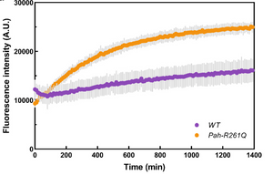
Bioinformatics analysis of PAH amyloidogenesis properties hinted that PAH has predicted a high (>50%) propensity to form intermolecular cross-β (amyloid-like) aggregates. To confirm their suspicions, the researchers studied amyloid-like aggregates formation by the mutant R261Q-PAH using Amytracker. A fluorescence assay with purified recombinant mutant protein compared to Wt protein mutant showed increased intensity over time indicating amyloid-like aggregation accumulation over time (5h). (Image: Amytracker 680 assay with recombinant purified proteins WT-PAH (purple) and p.R261Q-PAH (ochre), with 1 mg/ml of each protein in 20 mM Na-Hepes, 200 mM NaCl, pH 7 and incubation at 37 °C. Image from Supplementary Figure 6A by Aubi, O., Prestgård, K.S., Jung-KC, K. et al. (2021) Nature Communications, 12(1), 2073 (CC BY 4.0)).
This publication reports the complex nature of PKU, as loss-of-function mutation can be accompanied by toxic gain-of-function (aggregation & oxidative stress) for specific PKU-associated mutations. Amytracker was useful to characterize the formation of amyloid-like structures by the R261Q-PAH mutant protein.
Read More:
The structural basis of TDP-43 aggregation
Neurodegenerative diseases often involve the accumulation of misfolded proteins, that cells fail to refold or degrade and that eventually form aggregates. Among these, oligomers, which are small, still-soluble aggregates, are considered the most toxic and can lead to neuronal death. Identifying the aggregation-prone regions of these oligomer forming proteins is key for developing new therapeutic approaches.
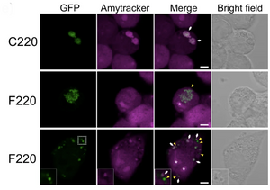
In over 97% of amyotrophic lateral sclerosis (ALS) cases, TDP-43 (TAR DNA/RNA-binding protein 43) condensates accumulate in the cytoplasm. The carboxy-terminal region of TDP-43 is processed into smaller aggregation-prone fragments that are included into these condensates. Akira Kitamura and their colleagues at Hokkaido University explored these fragments by expressing TDP-43 deletion constructs in murine neuroblastoma cells (N2A) and C. elegans. They found that TDP25, a fragment containing a glycine-rich domain and part of the RRM2 RNA-binding domain, is necessary and sufficient for condensate formation. The glycine-rich region drives this process, while the remaining N-terminal fragment is highly aggregation-prone. Only some of these condensates were stained by Amytracker 680, and therefore had an amyloid-like structure. This suggests that multivalent interactions and conformational changes in biomolecular condensates might facilitate the formation of amyloid aggregates of TDP-43. Expression of TDP25 caused cell death in N2A cells and reduced lifespan in C. elegans, suggesting this fragment contributes to the neurotoxicity observed in ALS. Therefore, stabilising TDP25 could be a potential therapeutic target to prevent toxic TDP-43 oligomers.
Image: Confocal images of N2A cells expressing different GFP-tagged TDP-43 carboxy-terminal fragments (green) stained with Amytracker 680 (magenta). In cells expressing the F220 fragment, not all the condensates formed are positive for Amytracker staining, suggesting that only a fraction of them contains amyloid fibrils. White arrows and yellow arrowheads represent the positions of Amytracker-positive and negative condensates in the cytoplasm. The asterisk in the images represents the nucleolus. Scale bar = 5 μm. Image from Figure 3E by Kitamura et al. (2024) Communications Biology Chemistry, 7(1), 743 (CC BY 4.0)
Read More:
Uncovering a new link to amyloid formation in neurodegenerative diseases
The 14-3-3 proteins regulate various cellular functions, including enzymatic activity and protein stability. The 14-3-3ζ isoform has been linked to neurodegenerative diseases due to its interaction with proteins like tau and α-synuclein, which form amyloid fibrils in Alzheimer’s and Parkinson’s. However, its direct role in amyloid plaque formation remains unclear.
A group of researchers from the Institute of Biotechnology in Vilnius aimed to determine if 14-3-3ζ can form amyloid fibrils. Using bioinformatic tools, they identified several aggregation-prone regions. To test amyloid formation under physiological conditions, they incubated 14-3-3ζ and monitored the process using amyloid-specific dyes: Thioflavin T, Congo Red, and Amytracker 630. Amytracker 630, which binds to amyloid fibrils, showed a significant increase in fluorescence, indicating the aggregation of 14-3-3ζ into amyloid-like structures. Additional methods, including FTIR, circular dichroism, and AFM, confirmed the structural transition from a helical structure to β-sheet-rich amyloid fibrils.
The study demonstrated that 14-3-3ζ forms amyloid fibrils, suggesting it may contribute to amyloid pathology in neurodegenerative diseases. This finding opens up new avenues for exploring the role of 14-3-3 proteins in neuropathies and highlights potential therapeutic strategies targeting this protein. The researchers also found that 14-3-3ζ amyloid formation reduced cell viability by 65% in neuroblastoma assays, indicating its neurotoxic effects.
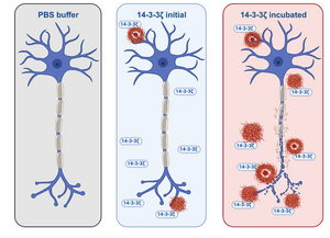
Image: Amytracker 630, a dye that binds to protein aggregates, showed a slight increase in fluorescence upon the initial addition of 14-3-3ζ protein. However, a significant rise in fluorescence was observed during the incubation period, suggesting that 14-3-3ζ forms amyloid fibrils over time. Image created with BioRender.
Read More:
Modular protein hydrogels for flexible applications
Hydrogels can be used as a 3D scaffold for cells to grow in tissue models and biofabrication applications. Such hydrogels can be made from proteins that form amyloid fibrils. Different biological applications have distinct requirements for the properties of these hydrogels. Designing a unique biopolymer for each application is cumbersome and to be able to assemble tailored systems from a set of predefined building blocks is beneficial.
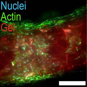
In a study, published in Acta Biomaterialia, researchers from ETH Zurich in Switzerland present an approach for a modular protein hydrogel platform. Their system allows the differentiation of hMSCs into osteoblasts, and the growth of neurite networks from primary neurons. In addition, their hydrogel system is compatible with 3D-printing of complex scaffolds. Self-assembly of these hydrogels from recombinant proteins happens within 1 h at a temperature of 60 °C.
Using Amytracker, the authors confirmed the assembly of the hydrogel from amyloid fibrils. They produced hydrogels from two types of building blocks (block A and block B). Block A is designed to form the amyloid fibrils and to facilitate the assembly of the polymer into a hydrogel. Block B contains bioactive amino acid motifs that can be modified if necessary. In this study RGD cell-attachment motifs were the motifs of choice. Block B at the same time acts as a flexible linker by connecting blocks A. The packing pattern and hardness of the hydrogel can be adjusted by varying the molar ratio between blocks A and B and by changing the length of block B.
Amyloid formation in ALS: Redefining the role of TDP-43 inclusions
Researchers from the University of Florence investigated the nature of TAR DNA-binding protein 43 (TDP-43) cytoplasmic inclusions, which are key pathological markers in neurodegenerative diseases like amyotrophic lateral sclerosis (ALS) and frontotemporal lobar degeneration (FTLD). The study focused on whether these inclusions exhibit amyloid-like characteristics, as there has been debate over their structure and classification.
The researchers conducted in situ and in vitro experiments to study TDP-43 inclusions. They expressed human TDP-43 in a motor neuron cell model and used Raman spectroscopy, FTIR, and TEM to examine aggregate structures. Purified TDP-43 was tested for amyloid formation using Amytracker 630.
The findings suggest that TDP-43 inclusions do not have amyloid characteristics, challenging the idea that they form amyloid-like structures. Instead, they likely form through a different mechanism.
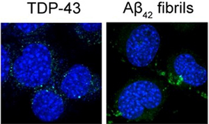
Image: Amytracker 630 was used to stain β fibrils, but it did not stain the cytoplasmic inclusions of TDP-43. Image from Figure 2 by Cascella, R. et al. (2023) Annals of Medicine, 55(1), 72-88 (CC BY 4.0).
Read More:
Amytracker confirms amyloid nature of inclusion bodies in muscle linked to gene mutations
Distal myopathies are genetically heterogeneous diseases that primarily affect skeletal muscles, particularly in the hands and feet, although they can progress to other muscles. Previous research has identified more than 25 genes associated with these conditions, but many patients still remain undiagnosed. SMPX (small muscle protein X-linked), a gene involved in muscle function, has previously been associated with non-muscle-related conditions such as hearing loss, but a study from Folkhälsan Research Center in Finland reveals its role in distal myopathy with protein inclusions.
The study, published in Acta Neuropathologica performed deep phenotyping and genetic sequencing on 10 patients from 9 families, revealing 4 distinct SMPX mutations, which cause progressive distal myopathy. By taking muscle biopsies and staining them with Amytracker 680, they confirmed that some of the sarcoplasmic inclusions present in the muscle fibres had amyloid-like characteristics. Moreover, cell culture experiments showed that these mutations increase protein aggregation and slow down the clearance of stress granules.
The study concludes that missense mutations in SMPX lead to a previously unknown form of distal myopathy, hallmarked by inclusion bodies in the muscles. These sarcoplasmic inclusions have amyloid-like characteristics - as highlighted by Amytracker staining - and represent a novel disease mechanism distinct from the hearing loss caused by other SMPX mutations.
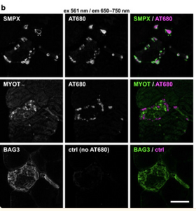
Image: Amyloid-like protein inclusions of SMPX in muscle tissue stained with Amytracker 680 (magenta) and SMPX and myotilin (green). Absence of Amytracker staining in the control (ctrl) sample confirms lack of autofluorescence from protein inclusions. Image from Figure 5B, Johari, M. et al. (2021) Acta Neuropathologica, 142, 375-393 (CC BY 4.0).
Read More:
Estrogen might be women’s secret weapon!
Alzheimer's disease (AD) affects women more than men. However, we don’t really know why. Women tend to live longer, which may contribute to a higher incidence of AD, but other factors, such as hormonal changes during menopause, could also influence disease progression.
In a study, published in Translational Psychiatry, researchers from the Centre for Brain Research in Bangalore explored why females exhibit a delayed onset of AD-related cognitive impairments how hormonal changes impact this progression.
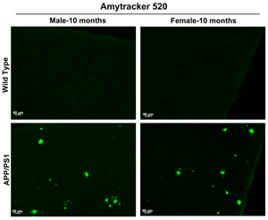
The researchers used male and female APP/PS1 transgenic mice which present with an AD phenotype relatively early in life to examine how synaptic protein translation changes over time in response to AD pathology. They also explored the role of estrogen in protecting against cognitive decline, by removing the ovaries from some of the female mice leading to abolished estrogen production. They used Amytracker 520 to track and compare the accumulation of amyloid plaques, a hallmark of AD, in male and female mice.
Interestingly, the researchers found that females with intact ovaries show slower cognitive decline, suggesting that estrogen plays a neuroprotective role. To support these assumptions, they analysed human data to assess sex differences in cognitive decline among people with familial AD and found that AD risk increases in women with menopause-related hormonal changes.
Image: Amytracker 520 (in green) selectively stains Aβ plaques in the brains of 10-month-old APP/PS1 mice, but shows no staining in wild-type mice. Image from Supplementary Figure 4 by Kommaddi, R.P. et al. (2023) Translational Psychiatry, 13, 123 (CC BY 4.0).
Read More:
Transition to aggregation in hnRNPA1A condensates visualised using Amytracker
Amyotrophic lateral sclerosis (ALS) and frontotemporal dementia are associated with the aggregation of certain RNA-binding proteins that are part of stress granules, including TAR DNA-binding protein 43 (TDP-43), Fused in Sarcoma (FUS) and Heterogeneous nuclear ribonucleoprotein A (hnRNPA). Stress granules are cytoplasmic membraneless organelles that are formed in response to stresses that halt protein production. These organelles collect the mRNA that was being translated, possibly to protect the RNA or to ensure that no damaged proteins are produced.
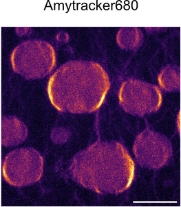
A study by Morelli et al. published in Nature Chemistry investigates how interactions with RNA modulate the aggregation of hnRNPA1A. Using an in vitro model around recombinant hnRNPA1A, the researchers show that this protein can phase-separate into molecular condensates where it then aggregates over time. Using Amytracker 680 to visualise hnRNPA1A aggregation, the authors could confirm that the amyloid fibrils are mainly found at the condensates’ surface.
Adding various concentrations of RNA into the system changes the dynamics of the condensation-to-aggregation process: In a system without RNA, hnRNPA1A condensates and progressively forms amyloid-like aggregates. However, this process is slow and does not reach a plateau in the timeline that was explored in the study. When RNA is added, hnRNPA1A aggregates faster, reaching a plateau before the end of the experiment and high amounts of RNA prevent condensation, but not aggregation, and hnRNPA1A forms aggregates directly.
Image: Confocal microscopy of hnRNPA1A condensates stained with Amytracker. reveal elevated fluorescence on the condensate surface indication transition from condenstation to aggregation of hnRNPA1A. Scale bar: 5 µm. Image from Figure S11 by Morelli, C., Faltova, L., Capasso Palmiero, U. et al. (2024) Nature Chemistry, 16(7), 1052-1061 (CC BY 4.0).
Read More:
Amytracker shows how α-Synuclein aggregates at the mitochondrial membrane
Faulty α-synuclein (α-Syn) aggregates in cells forming toxic α-Syn oligomers and finally Lewy Bodies which are the pathological hallmark of synucleinopathies. While Lewy Bodies are relatively easy to discern in a microscope, it turns out to be very difficult to investigate the early-intermediate forms of aggregates.
A study published in the journal Nature Neuroscience presents an assay to gain insight on the early events of α-Syn aggregation. In their paper Choi et al. make use of a phenomenon called Förster resonance energy transfer (FRET) which can assess to detect if light sensitive molecules are in very close contact to each other; because only then, the resonance energy can be transferred from a resonance energy donor to an acceptor. With modern microscopy techniques, scientists can excite a pair of fluorophores and detect FRET at a single-molecule level (smFRET), effectively turning FRET into a “spectroscopic ruler” to discern information about the conformational dynamics of various biomolecules, or the intramolecular distances amongst their building blocks.
Choi et al. make use of the smFRET technology by labelling mutated α-Syn with fluorescent tags acting as resonance energy donor/acceptor pair so that fluorescence emitted by the resonance acceptor, can be used as a proxy for α-Syn aggregation. They show that several α-Syn mutants show a concentration and time-dependent increase in the FRET signal and are able to track the structural changes of α-Syn from monomer, loose oligomers to dense higher FRET-efficiency oligomers. While monomers-oligomers appeared within hours, high-FRET oligomers only appeared after several days of incubation.
Correlating their fluorescence images with electron-micrographs by using Correlative Light Electron Microscopy, the researchers were able to show that the seeding events for α-Syn aggregation preferentially occur at the mitochondrial membrane. They further confirmed this result by showing that the signal from Amytracker co-localized with the signal of a fluorescent probe targeting cardiolipin present in the mitochondrial membrane.
In a neuronal cell line, they used Amytracker to confirm that α-Syn interacts with cardiolipin. As shown in their in vitro experiments, this interaction facilitates α-Syn aggregation, and traps cardiolipin in the growing aggregates. The accumulation of α-Syn aggregates on the mitochondria and the loss of cardiolipin can lead to an impairment of mitochondrial bioenergetics and induce mitochondrial dysfunction, which has been identified to be a major source of toxicity and the primary cause of neurodegeneration.
Image: In confocal images from neuronal cells, α-Synyclein aggregates are labelled with Amytracker 540 (red) and are shown to be in close contact with cardiolipin in the mitochondrial membrane (green). Image from Extended Data Figure 7 by Choi, M.L., Chappard, A., Singh, B.P. et al. (2022) Nature Neuroscience, 25, 1134-1148 (CC-BY-4.0).
Read More:
Studying the relationship between morphology and inflammatory effect of ɑ-synuclein aggregates
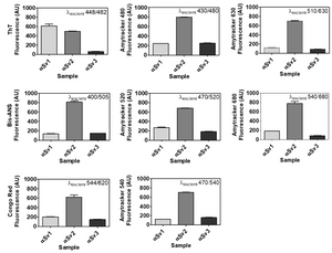
Parkinson's disease (PD) and Multiple System Atrophy (MSA) are both characterized by accumulation and misfolding of ɑ-synuclein (ɑ-syn). However, there are distinct differences between their pathology likely relating back to different strains of ɑ-syn aggregates. The goal of a study by De Luca et al. from IRCCS and SISSA in Italy was to analyze how neuronal SH-SY5Y cells reacted to different forms of ɑ-syn aggregates isolated from the olfactory mucosa of patients with PD and MSA as well as healthy controls using a Seed Amplification Assay. While ɑ-syn isolates from PD and MSA patients showed an efficient seeding activity, those from healthy controls did not. Seeded aggregates were labeled with an MJFR anti-alpha-synuclein aggregate antibody and showed that there is a certain structural difference between aggregates seeded with ɑ-syn isolated from PD and MSA olfactory mucosa, also confirmed by different grades of resistances to proteinase K. Analyzing a panel of inflammatory molecules, the researchers found that the inflammatory response was based on TLR2 activation and that the inflammatory response caused by MSA seeded aggregates was more pronounced. To better understand and analyze the structural differences between different ɑ-syn aggregate strains, the researchers generated ɑ-syn aggregate strains by fibrillating recombinant ɑ-syn under different conditions (ɑSv1, ɑSv2, ɑSv3). Structural differences in recombinant ɑ-syn strains were confirmed by Proteinase K treatment, MJFR antibody staining as well as a dye binding assay using Thioflavin T, Bis-ANS, CongoRed and all Amytrackter variants.
Image: Dye-binding assay of recombinant α-Syn aggregate strains. Once generated, αSv1, αSv2 and αSv3 were incubated with ThT, Bis-ANS, Congo red, Amytracker 480, Amytracker 520, Amytracker 540, Amytracker 630, Amytracker 680. αSv1 interacted with higher efficiency with ThT than αSv2 and αSv3. αSv2 interacted with higher efficiency with all the other fluorophores while αSv3 weakly interacted with all the fluorescent probes. Image from Figure S1 by De Luca et al. (2022) Cells, 11(1), 87 (CC BY 4.0).
Although capable to efficiently seed aggregation, the recombinant ɑ-syn strains did not transmit their seed-specific properties to the reaction products, which showed comparable biochemical properties. However, when used to stimulate SH-SY5Y cells, αSv1, αSv2, and αSv3 acted on different activators of inflammatory pathways, thus strengthening the existence of a correlation between morphological and inflammatory properties of αSyn fibrils.
Read More:
Faulty ribosome quality control triggers amyloid aggregation
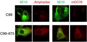
What causes the accumulation of toxic amyloid aggregates? An article by the group of Bingwei Lu from the Department of Pathology at Stanford University School of Medicine published in the journal Acta Neuropathologica Communications might have found the answer to this question. Their work shows that a defective ribosome quality control (RQC) causes the C-terminal fragment (APP.C99) of the Amyloid precursor protein (APP) to accumulate. This might be one of the earliest pathogenic events of amyloid aggregation. Labelling such aggregates in HeLa cells with Amytracker 680 shows that they have amyloid-like properties.
Image: Ribosome stall-induced APP.C99 with C-terminal extensions seeds amyloid ß-42 aggregation in HeLa cells. Fluorescent images showing aggregation of Aß42 (labeled by 6E10 antibody, green) as detected by Amytracker (left, red) as well as mOC78 antibody (right, red).Image from Figure S6F by Rimal, S. et al. (2021) Acta Neuropathologica Communications, 9, 169 (CC BY 4.0)
Read More:
Amytracker labels amyloid forms of tau in a cell based model for tauopathies
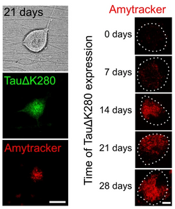
Accumulation of abnormal tau protein in the brain has been described as pathological hallmark of a group of neurodegenerative disorders called tauopathies. The most common tauopathy is Alzheimer’s disease (AD). In AD, intracellular inclusions containing aggregates of hyperphosphorylated tau coexist with extracellular amyloid plaques containing aggregated amyloid-β (Aβ). Clinically, tau inclusions correlate with cognitive impairment and therefore tau pathology is considered to be the major driver for neuronal degeneration and has become a target of rapidly evolving therapeutic strategies. Tau plays an important physiological function in dynamically interacting with axonal microtubules and is the target of various post-translational modifications. It is crucial that therapeutic agents conserve, or even restore the physiological function of tau, and prevent propagation of a range of abnormal forms of tau including soluble oligomers. Cell-based models of tauopathies are in high demand, as a platform for monitoring tau aggregation testing and efficacy of therapeutic compounds. A study published in the journal Nature Communications by Pinzi et al., describes a live-cell imaging assay to identify compounds that restore physiological microtubule interaction of an aggregation-prone full-length human tau construct in axon-like processes of model neurons and axons of primary neurons. The researchers used viral a single amino acid deletion mutant TauΔK280 of the tau gene which showed greater than 50% increased aggregation compared to wild-type tau in a heparin-induced cell-free aggregation assay to create a construct with an N-terminal photoactivatable GFP (PAGFP) reporter sequence. They successfully transfected neuronal cells (PC12) and primary neurons (DRG) with a viral vector and showed reduced interaction with microtubules of mutant TauΔK280 in the cellular environment using a technique called fluorescence decay after photoactivation (FDAP), likely due to oligomer formation. While initially, no amyloid forms of tau were identified in the cells using Amytracker 680, cells that were imaged 3 weeks after transfection showed accumulation of amyloid tau and Amytracker staining of cell bodies over time indicate significant formation of tau amyloids after 14 days of transfection.
After establishing their cellular model, the researchers proceeded to test the efficacy of potential therapeutic agents preventing tau aggregation. A compound called PHOX15 was able to restore mutant TauΔK280 interaction with microtubules, likely by preventing oligomer formation. While PHOX15 was not able to reverse aggregation, once amyloids were present, it was able to reduce the formation of tau amyloids in neurons. Taken together, the researchers showed that PHOX15 reduces the formation of tau filaments and decreases the phosphorylation of tau at disease-relevant sites thereby restoring the interaction of tau with microtubules to a physiological level. Their cell based model for testing and careful characterization of the compound distinguish PHOX15 as a promising therapeutic target and may advance therapeutic approaches that specifically target the key traits of tauopathies.
Image: Presence of tau amyloids in DRG neurons. Left: PAGFP-TauΔK280 and Amytracker 680 fluorescence indicate formation of tau amyloids in the neuronal cell body after 21 days (scale bar: 20 µm). Right: Amytracker 680 signal indicates the formation of tau amyloids in the neuronal cell body on day 0-28 (scale bar: 20 µm). Image from Figure 2DE by Pinzi, L. et al. (2024) Nature Communications, 15(1), 1679 (CC BY 4.0).
Read More:
Amyloids trigger proinflammatory signalling Multiple Myeloma
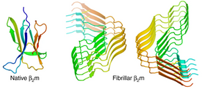
Multiple myeloma is a complex B-cell malignancy characterised by the accumulation of malignant plasma cells. Progression of multiple myeloma is related to dysregulated inflammatiory processes; especially leucocytes and tumor-associated macrophages (TAMs) are central during the initiation and progression of the disease and high concentration of TAMs often relates to drug resistance and fast proliferation. TAMs produce proinflammatory cytokines whose production is controlled carefully in the cell under normal circumstances. The production and release of proinflammatory cytokines is controlled by nod-like receptor NLRP3. While the activation process of NLRP3 is poorly understood, it has been found that, among other triggers, endogenous amyloid fibrils activate NLRP3. In a study published in the journal Immunity, Hofbauer et al. from University Hospital Erlangen in Germany aimed to understand the role of amyloid fibrils in the activation of NLRP3. For this, they used Beta-2-microglobulin (b2m) which is well known for amyloid aggregation propensity. B2m is a small non-glycosylated protein and universally expressed in all nucleated cells. Interestingly, b2m concentration is known to increase in myelomas and is correlated with a poor prognosis. However, the molecular mechanism behind this is not known. The researchers used Amytracker 480 to verify the formation of b2m amyloids in macrophages. Since Amytracker 480 is non-toxic and cell-permeant (Macrophages were washed in PBS and incubated with Amytracker 480 (1:1000) for 1h at 37 °C), amyloid presence in macrophages was quantified using flow cytometry and Amytracker fluorescence was used as an indicator for presence of amyloids. The researchers found that the NLRP3 inflammasome is activated after phagocytosis of β2m and that internalised β2m aggregates into amyloid fibrils under the acidic phagosomal conditions, which results in lysosomal swelling and damage. They further demonstrate that the β2m-triggered NLRP3 activation in TAMs results in the release of proinflammatory cytokines and in turn favours the growth and severity of Multiple Myeloma. These findings provide a strong indication that dysregulated inflammatory response is a driver of disease in Multiple Myeloma and that inhibition of NLRP3 represents a potential therapeutic approach.
Image: The structure of β-2-microglobulin in its native and fibrillar states. Image from Figure 4 AB by Iadanza, M.G., Silvers, R., Boardman, J. et al. (2018) Nature Communications, 9(1), 4517 (CC BY 4.0).
Read More:
Amytracker: a tool to identify toxic dipeptide repeats
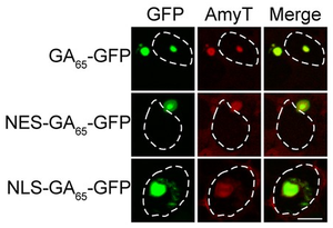
Repetitive genomic regions are known to expand across generations, due to errors during their replication, and cause a series of - mainly - neurological diseases collectively called repeat expansion diseases. One of these repetitive genomic regions is the GGGGCC (G4C2) locus in the C9orf72 gene. Its expansion is linked with amyotrophic lateral sclerosis (ALS) and frontotemporal dementia (FTD). It is still unclear, however, whether the cytotoxic effects of the repeat-expansion are emerging from the G4C2 mRNA, or from the results of its translation. Frederic Frottin from F. Ulrich Hartl’s group at the Max Planck Institute of Biochemistry and Mark S Hipp at the University Medical Center Groningen tried to dissect the issue in their paper published in the eLife journal. In order to distinguish the effects of the mRNA from the ones of the resulting protein, the researchers designed a construct that produces a poly-GA containing protein from a non-G4C2 RNA.They then used a Nuclear Export Signal (NES) and Nuclear Localization Signal (NLS) to localise the construct on either side of the nuclear envelope. Both constructs formed inclusions in their respective compartments which were stained by Amytracker 680, confirming the presence of amyloid structures within them. Leveraging this system, the authors were able to shed light on the toxic effects of the G4C2 expansion in the C9orf72 gene. Their work reveals that the expression of cytoplasmic poly-GA has only modest effects on cell viability, and that nuclear poly-GA proteins, however, are more toxic and impair nucleolar protein quality control and protein biosynthesis.
Image: Cytoplasmic and nuclear aggregates in HEK293 cells stained with Amytracker 680 (AmyT, red). Poly-GA DPRs were visualised by GFP fluorescence (green). White dashed lines delineate the nucleus based on DAPI staining. Image from Figure 1D by Frottin, F. et al. (2021) eLife, 10, e62718 (CC BY 4.0).
Read More:
How mutations contribute to pathological diversity in synucleopathies
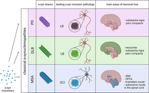
Alpha-synuclein (α-Syn) is a presynaptic neuronal protein encoded by the SNCA gene. It is expressed heavily in the brain and regulates synaptic vesicle trafficking and subsequent neurotransmitter release. Under certain conditions, α-Syn aggregates and forms, together with other components, intracellular inclusions called Lewy bodies (LBs) or Lewy Neurites (LN), which are the defining pathological hallmark of many synucleinopathies. Several familial mutations of the SNCA gene have been identified causing LB and LN pathology, but manifest in different diseases like Parkinson's Disease, Dementia with Lewy bodies (DLB) or Multiple System Atrophy (MSA), suggesting that the various mutations may act via distinct mechanisms.
Image: Various pathologies like Parkinson’s Disease (PD), Dementia with Lewy Bodies (DLB) and Multiple Systems Atrophy (MSA) are caused by different α-Syn strains. Image from Figure 1 by Malfertheiner, K. et al. (2021) Frontiers in Neurology, 12, 737195 (CC BY).
In a study published in Science Advances, Senthilt Kumar and colleagues from Hilal A. Lashuel’s group at the EPFL, Switzerland, provided an in-depth characterization of a novel SNCA mutation which is unique as it is the first one that is located in the Non-Amyloid Component (NAC) domain. The patient carrying this mutation presented with DLB and atypical frontotemporal lobar degeneration. The researchers produced recombinant wild-type (wt) and mutant E83Q α-Syn and showed that the E83Q mutation accelerates the aggregation kinetics of α-Syn and that fibrils of α-Syn show distinct morphology, stability, and structural features in vitro. Since α-Syn is a membrane protein the researchers wondered if the E83Q mutation influences membrane interaction. Using artificial vesicle and cellular models they show that the E83Q mutation does not strongly alter α-Syn binding to lipid vesicles or intracellular membranes and does not promote the formation of pathological α-Syn aggregates in mammalian cell lines. However, a greater propensity of E83Q α-Syn to form cytotoxic oligomers and higher seeding activity was established. Interestingly, the researchers were able to show that seeding with E83Q α-Syn preformed fibrils, but not WT α-Syn preformed fibrils leads to formation of Lewy Body-like inclusions in neurons. While classical markers were used to characterise the protein components, Amytracker was valuable to determine the amyloid character within these inclusions. Taken together, the study by Kumar et al. provides an impressively complete picture on how a single mutation in the SNCA gene can unlock the pathogenicity of human α-Syn fibrils and showcases how the pathological diversity of synucleinopathies might be related to specific strains of α-Syn fibrils.
Read More:
Deciphering the complexity of intracellular Tau aggregates
Tau is an intrinsically disordered protein with the key function to stabilise axonal microtubules. In several neurological disorders collectively known as Tauopathies, tau proteins form aggregates that accumulate in cells and cause neuronal damage. The molecular mechanism which leads to tau aggregation is complex and liquid–liquid phase separation (LLPS), a process that is used by cells to assemble membrane‐less organelles, has been found to be involved in Tau aggregation. De-mixing of Tau can be triggered through the addition of macromolecular crowding agents, or through coacervation (i.e. co-condensation of the positively charged Tau microtubule‐assembly domain with polyanionic RNA into liquid‐like droplets). In the complex ecosystem of the neuronal cytoplasm, it is possible that Tau undergoes crowding and coacervation at the same time. To investigate this scenario, researchers around Susanne Wegmann at DZNE in Berlin, Germany conducted a study investigating the effect of molecular crowding and RNA interaction on Tau aggregation in cells. These findings from Hochmair et al. were published in the EMBO Journal. Using an in vitro model, they find that molecular crowding is necessary to enable Tau and phosphorylated Tau coacervation with RNA. When the researchers treated HEK sensor cells with Tau:RNA condensates, they observed three categories of intracellular aggregates: CYT-Tau: large, bright cytoplasmic inclusions, NUC-Tau: often bright and nuclear, and NE-Tau: dimmer inclusions that reside at the nuclear envelope and form a thin non-uniform layer of small granule like structures. The researchers used FRAP (Fluorescence Recovery After Photobleaching) and FLIM (Fluorescence lifetime imaging) to investigate differences between the intracellular aggregate and found that NUC-Tau is the most dense followed by CYT-Tau and NE-Tau. Interestingly, Amytracker 680 labels CYT-Tau and NUC-Tau, but not NE-Tau confirming that these three types of aggregates have different morphology and molecular content. When they used Alzheimer’s Disease brain lysate instead of in-vitro prepared Tau:RNA condensate to treat HEK cells, the researchers were able to recapitulate these findings. Taken together, the study by Hochmair et al. contributes to deciphering the complexity of Tau aggregation and explains the variety of disease‐related cellular Tau accumulations.
Image: Amytrackers labels bona-fide intracellular aggregates in cortical tissue sections showing pronounced AD pathology and reveals a clear fibrillar morphology assembled in tangled net-like structures. ©Ebba Biotech
Read More:
Linking aggregate size and toxicity using Amytracker
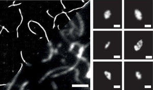
In the context of neurodegenerative diseases, large, fibrillar amyloid deposits - such as Lewy bodies, Amyloid plaques, and Neurofibrillary tangles - characterize the pathology of the disease but it has recently become evident that small, spherical aggregates below 20 nm in diameter are mainly responsible of the toxic response that results in neurodegeneration. The small size of these aggregates makes their detection difficult, especially within biologically relevant environments. To tackle this issue, Michael Morten and their colleagues at the Imperial College in London have developed a method for single-molecule localization microscopy (SMLM) using aggregate-activated fluorophores. Initially, they benchmarked Alexa647, Thioflavin T, Proteo-Stat and Amytracker 630 staining of aggregates assembled from recombinant α-Synuclein (α-Syn) using standard TIRF imaging. Since Thioflavin T lacked in brightness, it was imaged with 6x more laser power (60 mW) compared to the other fluorophores (10 mW). Exploring the relationship between the total fluorescence intensity detected from each single aggregate versus its size, the researchers found that Proteo-Stat and Amytracker 630 demonstrated greater fluorescence intensities with aggregate size compared to Thioflavin T and even Alexa647, which is due to higher densities of fluorophores bound to each aggregate. This highlights the advantages of aggregate-activated fluorophores over the Alexa647-conjugation approach. Due to the Abbe diffraction limit, determining the size of aggregates <200 nm is challenging. Therefore, the researchers used SMLM to characterize the structural features of aggregates in finer detail. They found that Amytracker 630 is suitable for quantitative SMLM imaging of aggregates at circa 4 nm precision and enables sensitive and quantitative detection of aggregate species down to approximately 10 nm in size in live cells and ex vivo brain tissue.
Image: Amytracker 630 labeled recombinant α-Syn aggregates. Left: imaged by SMLM reconstruction and conventional diffraction-limited TIRF (2 µm scale bar). Right: Smaller aggregates detected using SMLM (0.1 µm scale bar). Image from Figure 2D by Morten, M.J. et al. (2022) PNAS 119(41), e2205591119 (CC BY 4.0)).
An advantage of using Amytracker 630 compared to the other fluorophores was its compatibility with live cell imaging. When incubated with recombinant α-Syn, Amytracker 630 staining of live HEK293A cells with GFP-tagged proteasomes revealed that the plasma membrane effectively prevents aggregates >450 ± 60 nm from entering the cell, and that internalized aggregates are quickly surrounded by proteasomes, suggesting that proteostasis mechanisms respond promptly to proteotoxicity. Finally, the size of the membrane-penetrating aggregates detected by Amytracker 630 was shown to correlate with cytotoxicity. When the cytotoxicity assay was performed with aggregates from recombinant α-Syn as well as brain derived aggregates from Parkinson’s Disease and Lewy Body Dementia, it was shown that aggregates differ in toxicity depending on the pathology they originated from. Taken together, the study by Morten et al. is an impressive example of technology development directly benefiting the medical field by providing important clues about the relationship between aggregate size and toxicity.
Read More:
Protein aggregation in wound healing
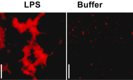
Lipopolysaccharide (LPS) is a cell envelope glycolipid produced by most gram-negative bacteria. When LPS is recognized by Toll-like receptor 4 (TLR4) an innate host-response to bacterial infection is triggered. To achieve a robust antibacterial response while maintaining control of inflammatory processes, LPS has to be cleared from the infection site. A group of researchers around Prof. Artur Schmidtchen from the Division of Dermatology and Venereology at Lund University have investigate the mechanism of LPS clearing from wounds and shown that addition of LPS or bacteria to acute wound fluid (AWF) leads to precipitation of proteins mediating LPS aggregation and scavenging. In a recent study, Petrlova et al. sought to define the LPS interactome in AWF and investigate the functional consequences of LPS aggregation. The researchers use Amytracker 680 to confirm the presence of protein aggregates in AWF treated with high concentrations of LPS (Image: Acute wound fluid incubated with LPS from E.coli or Buffer (negative control) labelled with Amytracker 680 and imaged using fluorescence microscopy. Image from Figure 2D, Petrlova, J. et al. (2023) iScience, 26(10), 107951 (CC BY 4.0).
Using Mass Spectroscopy and western blot, the researchers show that wound-fluid aggregates contain not only thrombin but also a multitude of other proteins, including coagulation factors, annexins, histones, antimicrobial proteins/ peptides, and apo-lipoproteins with many of them also being present in plaques from patients with systemic- or neurodegenerative amyloidoses. To study the functional effect of LPS induced aggregation in acute wound fluids, the researchers performed experiments with reporter monocytes and mice that produce a quantifiable response upon TLR4 activation. These experiments showed that wound fluid inhibits the inflammatory cascade induced by LPS, both in cells and in vivo. While presenting evidence of amyloid formation in a functional context, the study provides another puzzle piece linking amyloid formation to inflammation. Understanding the functional aspect of amyloid formation might pave the way for innovative strategies to mitigate the detrimental effects of amyloid diseases.
Read More:
Phase separated α-synuclein is more prone to aggregation
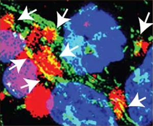
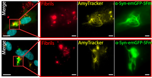
Alpha-synuclein (α-Syn) aggregation hallmarks a group of neurodegenerative diseases known as synucleinopathies with Parkinson’s disease as its most well-known representative. The molecular mechanism behind the aggregation of α-Syn is still not completely understood. Many aggregation-prone proteins, like α-Syn, are known to form phase-separated condensates and recent findings suggest that the early events that lead to protein aggregation, amyloid deposition, and ultimately neurodegeneration, are happening within these condensates. Recent work from Piroska et al. published in the journal Science Advances found strong evidence of a connection between phase separation and aggregation of α-Syn. When α-Syn was present in form of a fusion protein with GFP and fragments of a faulty chaperone that facilitates multivalent interactions and formation of phase separated condensates, treatment of SH-SY5Y cells with pre-formed fibrils (PFFs) leads to formation of solid structures forming needle-like protrusions within a few hours. These α-Syn condensates show the characteristics of amyloid deposits, as they are not dissolved by detergents and stop exchanging components with the surrounding cytoplasm. Moreover, they are intensely labelled by Amytracker 630, confirming the presence of amyloid structures within them, while α-Syn condensates formed before exposure to pre-formed fibrils are not. In other words, when α-Syn separates into condensates it is more prone to aggregation and this is probably due to the elevated local concentration of α-Syn within these aggregates. Linking supersaturation of α-Syn in phase-separated compartments with amyloid formation gives important insights into the early stages of neurodegenerative conditions and might provide clues on how to interfere with preventative measures.
Image: Pre-formed fibrils trigger the evolution of α-Syn condensates into solid-needle-like structures in SH-SY5Y neuronal cells. Nuclei (cyan), pre-formed α-Syn fibrils (red), Amytracker (yellow), and α-Syn-GFP (green). Image from Figure S6B, Piroska, L. et al. (2023) Science Advances, 9(33), eadg5663 (CC-BY-4.0).
Read More:
FL-OPTIR for studying biophysical properties of amyloids in cells and tissues
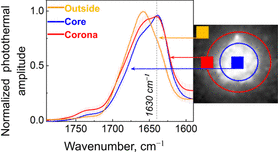
Infrared (IR) spectroscopy is an invaluable tool to study the biophysical properties of amyloid aggregates. Although historically used as a tool for composition analysis of chemical compounds as it measures the vibrational energy of chemical bonds, IR spectroscopy provides measures to analyse the size and rigidity of amyloid fibrils. Coupling of IR spectroscopy to an optical microscope (OPTIR) is a new method that allows users to obtain chemical information of biological macromolecules from cells and tissues at submicron resolution. Using OPTIR for investigating the biophysical properties of amyloid fibrils in cells and tissues has been challenging since amyloid fibrils cannot be easily identified by conventional light microscopy. Therefore, Oxana Klementieva and her research group at Lund University in Sweden combined epifluorescence imaging and OPTIR in a single instrument. Their “Fluorescence-Located” OPTIR or FL-OPTIR allows precise targeting using epifluorescence signals to locate amyloid-rich regions for OPTIR analysis. In a recent study published in Journal of Medicinal Chemistry, Prater et al. used fixed sections of APP/PS1 transgenic mouse brain and labelled amyloids using Amytracker 520. Guiding OPTIR measurements via the fluorescent signal from Amytracker 520, detailed biophysical analysis of amyloids in the core, corona, and outside of the plaque could be performed indicating a high degree of heterogeneity in the distribution of β-sheet structures within the amyloid plaque. Therefore, the FL-OPTIR method with the optotracer Amytracker might be valuable in studying amyloids in cells or tissue.
Image: FL-OPTIR spectra recorded from the outside, core, and plaque corona as indicated by the markers of the corresponding colour on the inset representing an amyloid plaque stained with Amytracker 520. Image from Figure 2D, Prater, C. et al. (2023) Journal of Medicinal Chemistry 66(4), 2542–2549 (CC BY 4.0).
Read More:
Spatial pattern of microglial activation in relation to amyloid plaques
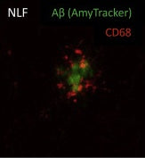
Microglia cells play a vital role in regulating brain development, maintenance of neuronal networks, and injury repair. Their involvement in the progression of protein aggregation leading to neurodegenerative diseases is complex. While they can clear amyloid plaques and prevent their accumulation, dysfunctional microglia regulation may be a key factor in disease progression. Microglia can “sense” accumulation of amyloid plaques which leads to a change in the expression patterns of Plaque Induced Genes (PIGs). One well-known PIG is Trem2, a microglial membrane receptor protein linked with Alzheimer’s disease (AD). A key function of Trem2 is to induce the microglia cells towards a phagocytic phenotype. Therefore, mutations in Trem2 lead to a higher risk of AD. In a study published by Wood et al. in Cell Reports, researchers set out to understand how Trem2 activation works and how it is related to the distance from a plaque. The researchers used spatial transcriptomics to investigate the spatial distribution of gene expression in NLF mice and NLF mice with R47H mutation in Trem2 which is considered a major risk factor for developing AD. To be able to define regions of interest for transcriptomics, Amytracker 520 was used to label plaques. Based on Amytracker 520 signal, radial regions were defined as “on-plaque”, “periplaque” or “away from plaque”. Based on this categorisation, the researchers were able to determine that, from a total of 55 PIGs, 23 (including Trem2) were only upregulated in “on plaque” regions. The significant expression differences between “on-plaque” and “away from plaque” regions observed in mice with functional Trem2 were not apparent in the mice with mutated Trem2. Looking into the functional aspect, the researchers found that, close to plaques, microglia cells express a higher level of CD68 indicative of a phagocytic character. This phenotype was, again, not apparent in mice with mutated Trem2 indicating that the phagocytic phenotypic change of microglia (i.e. CD68 expression) is dependent on the Trem2 genotype. The study highlights the importance of functional microglial activation via Trem2 and the spatial pattern of Trem2 related microglial activation in relation to plaque formation.
Image: Hippocampal section of PFA fixed NLF mouse brain, labelled with Amytracker 520 (green) for amyloid plaques and CD68 (red) indicating the phagocytic phenotype of microglial cells in close proximity to the amyloid plaque. Image from Figure 5C, Wood, J.I. et al. (2022) Cell Reports 41, 111686 (CC BY 4.0).
Read More:
Amyloidogenicity of SARS-CoV-2 spike protein
Infection with SARS-CoV-2 leads to patients developing the COVID-19 disease, which is a complex hyperinflammatory syndrome, characterised by acute respiratory distress (ARD). Aside from these severe respiratory symptoms, we now know that the virus can present in unexpected, varied, and long-lasting manners. Recent studies hint towards the amyloidogenicity of the SARS-CoV-2 spike protein (Nyström, S. et al. 2022) and indicate the presence of amyloid aggregates in plasma clots from patients suffering from Long-Covid symptoms (Pretorius, E. et al. 2021)
Researchers from the University of Lund in Sweden and the National University of Singapore previously found that spike protein binds to a range of hydrophobic molecules including bacterial lipopolysaccharide (LPS). This is a highly interesting finding considering that the Covid-19 disease is linked to hyperinflammation and LPS binding to pattern-recognition receptors, which play a key role in innate immunity, kickstarting inflammatory processes. The LPS binding sites on the spike protein were found adjacent to regions that are considered aggregation-prone, including the ones that are able to form amyloid structures, suggesting that LPS might modulate aggregation through these residues. In their most recent publication, Petrlova et al. wondered whether LPS contributes to the amyloidogenic properties of spike protein. Using available structures of dimers and trimers of spike protein, they simulated aggregation in presence of LPS and their results suggested that spike protein and lipid-A (a major component of LPS) can form stable higher-order complexes that the spike protein on its own cannot form. The researchers used Amytracker 680 to visualise aggregates formed by spike protein and found small and rounded aggregates (0.02-0.2 µm). When the spike protein was treated with LPS, these aggregates increased to form particles with up to 2 µm. Although, amyloid formation of spike protein and LPS warrants further investigation to study its relevance in vivo. However, understanding the link between spike protein aggregation and amyloid formation will have important implications for diagnostic and therapeutic approaches
Read More:
Other Resources:
Lewy body formation in seeding based neuronal models
Parkinson’s disease is a brain disorder that causes unintended or uncontrollable movements, such as shaking, stiffness, and difficulty with balance and coordination. Symptoms usually begin gradually and worsen over time. As the disease progresses, people may have difficulty walking and talking. The basis of these symptoms is progressive neurodegeneration and accumulation of degradation-resistant intracellular aggregates, termed Lewy bodies, throughout the brain.
One of the major components of Lewy bodies is α-synuclein, a small protein that normally localizes at the presynaptic terminal of neurons where it regulates the release of neurotransmitters and possibly participates in the assembly of the cytoskeleton. The main characteristic of α-synuclein is that it is not tightly folded like most other proteins, but some regions of it can adapt to the shape of its interaction partners. The intrinsically disordered structure of α-synuclein also makes it prone to aggregation. Aggregated α-synuclein disrupts mitochondrial function and therefore leads to neuronal death. Moreover, aggregates of α-synuclein can act in a prion-like manner by seeding further aggregations, and spreading from one region of the brain to others, partially explaining the progressiveness of Parkinson’s disease.
The molecular mechanism behind α-synuclein aggregation and its correlation with Lewy body formation and neurodegeneration is poorly understood, and appropriate models for studying α-synuclein aggregation are lacking. The most common model for α-synuclein fibrillization is a seeding-based approach in which formation of intracellular aggregates is induced by adding a small amount of α-synuclein fibrils to a neuronal culture. Normally, the cells are then kept in culture for 14 days and α-synuclein fibrillization is studied. Under these conditions, however, only few cells show inclusions that resemble Lewy bodies.
Researchers from the lab of Professor Hilal A. Lashuel at the Ecole Polytechnique Fédérale de Lausanne (EPFL) in Switzerland hypothesized that the maturation process of transitioning from α-synuclein fibrils to Lewy bodies might require a longer time than designated by the standard model. Therefore, the authors prolonged incubation time from 14 to 21 days. At this later time point, inclusions with both an immunocytochemical profile and morphological structure similar to bona fide Lewy bodies were observed in about 20% of the neurons.
To characterize the effects of α-synuclein seeding over time, the authors used an antibody to label phosphorylated α-synuclein (pS129) which is the predominant form in Lewy bodies. In addition, they used antibodies labeling proteins that are a common component in Lewy bodies, like α-synuclein as well as ubiquitin and ubiquitin binding protein (p62) to characterize the effect of α-synuclein seeding. Finally, they used Amytracker 680 to label structures containing repetitive β-sheets to confirm the amyloid-like nature of the seeded aggregates. Indeed, Amytracker 680 colocalized with phosphorylated α-synuclein at days 7, 14 and 21 after seeding. While the seeded aggregates shared the immunohistochemical profile of Lewy bodies already at an early stage, the morphological features of bona fide Lewy bodies like their association with various organelles, including mitochondria became evident only at a later stage. It was observed in a few cases after 14 days, but was much more evident after 21 days.
Due to the observed association of Lewy Body-like inclusions with mitochondria, the authors investigated how the association with Lewy bodies affects mitochondrial function. They found that 21 days after the seeding of α-synuclein, but not after 7 or 14 days, mitochondrial respiration was severely impaired confirming that the formation of Lewy bodies might be associated with mitochondrial dysfunction. Using transcriptomic analysis, the authors showed that the expression of genes that regulate neuronal cell death was significantly altered at the later time points of the study. Furthermore, α-synuclein mRNA levels were significantly reduced at day 21 suggesting that the neurons are trying to prevent any further aggregation.
In summary, the publication by Mahul-Mellier et al. makes a strong point for increasing incubation times in seeding based models to be able to study the processes leading to the maturation and formation of Lewy bodies, which can reveal new insights into the pathogenesis of Parkinson’s disease.
Read More:
Other Resources:
Prolonged stress leads to accumulation of misfolded proteins in the nucleolus
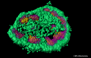
The nuclear proteome is rich in proteins which are prone to aggregate upon conformational stress. This might explain why intranuclear inclusions can often be found in neurodegenerative disorders associated with protein aggregation. Using a combination of fluorescence imaging, biochemical analyses, and proteomics, researchers at Max Planck Institute for Biochemistry around Prof. F.-Ulrich Hartl have investigated the role of the nucleolus as a “phase-separated protein quality control compartment” and published their results in the renowned scientific journal Science. The nucleolus is the largest non–membrane-bound nuclear subcompartment and consists of liquid-like phases that do not intermix, giving rise to distinct zones. To investigate the fate of a nuclear protein during heat stress, the researchers generated a cell line producing a reporter protein called NLS-LG. NLS-LG carries a nuclear localization signal, to make sure it is distributed to the nucleus. The protein's location can be tracked by a heat stable fluorescent signal and its folding state can be tracked with a thermolabile fluorescent signal. When the researchers exposed the cells to heat, they saw that NLS-LG was unfolded and transferred into the outermost zone of the nucleolus. (Figure: The outermost zone of the nucleolus is the granular component (GC) phase marked in green.) Upon recovery from heat stress, NLS-LG was again relocated to the nucleus and a bright fluorescent signal indicated successful refolding. It was further shown that Hsp70, which is an important part of the cell's machinery for protein folding, also transferred to the nucleolus upon heat stress. When the activity of Hsp70 was inhibited, relocation of NLS-LG to the nucleus after recovery from heat stress was also prevented.
Image: Super-resolution light microscopy shows that the nucleolus is not separated from the rest of the cell by a membrane and that it consists of different zones, which are distinct from each other and membrane-less as well.© MPI of Biochemistry)
When nucleolar organization was disrupted by a toxin that causes nucleolar disassembly and the cells were exposed to heat, instead of translocating to the nucleolus, the NLS-LG reporter protein formed aggregate foci in the nucleus. Using Amytracker 680, the authors were able to demonstrate that these nucleoplasmic foci, in contrast to nucleolar assemblies, were positive for amyloids with a cross β structure. Although recovery from heat stress was slow and inefficient in the nucleoplasmic foci, a certain capability of refolding was evident since Amytracker 680 fluorescence intensity decreased upon recovery from heat stress. This demonstrates that Amytracker 680 can also be used to label reversible aggregates on top of irreversible amyloid fibrils. To explore the protective capacity of nucleolar quality control, cells were exposed to prolonged heat stress. This led to an increase in nucleolar volume attributed to the influx of misfolded protein and to a transition in nucleolar appearance from liquid droplet-like to a hardened state. When Amytracker 680 was used under these conditions in cells expressing the NLS-LG reporter protein, a distinct nucleolar signal indicated presence of amyloids with a cross β structure, which dissolved slowly upon recovery from heat stress. This shows the potential of Amytracker 680 to report the presence of amyloids in phase separated compartments like nucleoli highlighting Amytracker as a tool to be used in in vitro liquid-liquid phase separation experiments for studying protein aggregation and amyloid formation.
Taken together, the publication by Frottin et al. pioneers the notion that “the nucleolus serves as a storage compartment for a subset of misfolded proteins under proteotoxic stress conditions, preserving them in a state competent for refolding or degradation”. The impairment of nucleolar quality control through environmental stressors and other factors might directly contribute to the emergence of idiopathic neurodegenerative pathology.
Read More:
Amytracker can be used for intracerebral multiphoton microscopy
A team of researchers from Massachusetts General Hospital and Harvard Medical School as well as Linköping University have used a fluorescent probe of the same type as Amytracker for multiphoton imaging of Amyloid-β deposits in transgenic mice in vivo.
The fluorescent molecule clearly targeted and labeled core plaques in the cerebral tissue and vasculature when imaged after intravenous injection. The fluorescent signal appeared already shortly after injection, however reaching maximal intensity between 24 and 72 hours – clearly showing the capacity of the probe to pass the blood brain barrier without causing toxicity. Since core plaques are labeled intensely with the fluorescent molecule, emitting in the red channel, the molecule can also be combined with other markers for diffuse plaques or neurofibrillary tangles with emission in the green or blue channel.
Read More:
The clue to detect multiple systems atrophy?
In a study, recently published in Nature, a fluorescent tracer molecule similar to Amytracker has been used to detect α-synuclein aggregates in cerebrospinal fluid from patients with synucleopathy. Interestingly, the fluorescent tracer molecule was binding aggregates from patients with Multiple Systems Atrophy with higher affinity than aggregates from patients with Parkinson’s Disease. While aggregates of α-synuclein in distinct synucleinopathies have been proposed to represent different conformational strains of α-synuclein, this is the first study that provides distinct clues that can be used in future diagnostic assays.
Read More:
Anomalous fibrin amyloid formation
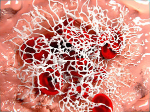
Lipopolysaccharides (LPS) from the Gram-negative cell envelope can be shed from dormant bacteria or from continual bacteria entry into the blood and serve to contribute to the chronic inflammation. The presence of highly substoichiometric amounts of LPS from Gram-negative bacteria caused fibrinogen clotting to lead to the formation of an amyloid form of fibrin. The teams around Prof. Douglas B. Kell from the University of Manchester and Prof. Etheresia Pretorius from the Stellenbosh University have shown that the broadly equivalent lipoteichoic acids (LTAs) from two species of Gram-positive bacteria have similarly (if not more) potent effects than LPS. Specifically they have showed that LPS, iron, and the 2 LTAs cause amyloid formation of plasma proteins, and in particular of fibrin(ogen) as blood is clotted. They confirmed amyloidogenesis by using Amytracker 480 and Amytracker 680 to identify amyloid protein deposits.
Read More:
Amyloids in type-2 diabetes
Type-2 diabetes is a progressive condition marked by resistance towards the blood-sugar regulating hormone insulin. Recently, Type-2 diabetes is become recognized as an inflammatory condition which is often accompanied by cardiovascular complications. The teams around Prof. Etheresia Pretorius from Stellenbosh University and Prof. Douglas B. Kell from the University of Manchester investigated blood clot formation from plasma of diabetic patients. Using advanced confocal microscopy and Structured Illumination Superresolution Microscopy, the authors found that plasma clots from diabetic patients contain significant amounts of amyloid hinting towards an inflammatory cue for the occurrence of cardiovascular symptoms in Type-2 diabetes.
Read More:
Advanced imaging
In a new study in the Journal of visualized experiments, the team around Prof. Peter Nilsson and Prof. Per Hammarström from Linköpings University describe how luminescent conjugated oligothiophenes (LCOs) can be used with Hyperspectral Imaging (HIS) and Fluorescence Lifetime Imaging (FLIM) to detect amyloid species. In a practical approach, the authors highlight caveats of LCO staining and give valuable advice on troubleshooting.
Read More:
Protein engineering for better PET radioligands
An approved method for visualization of Amyloid β (Aβ) plaques in patients suffering from Alzheimer's disease is Positron Emission Tomography (PET). This method requires a radiolabeled amyloid ligand. A frequently used molecule is Pittsburgh Compound B (PIB) which is a derivative of Thioflavin T. Radiolabeled PIB is well-suited to visualize insoluble Aβ plaques and has been used for differential diagnosis of AD. However, the PIB signal, giving an estimate of total plaque load becomes saturated early in the disease progression. To being able to diagnose Alzheimer's disease early-on, soluble forms of aggregated Aβ like oligomers and protofibrils need to be visualized
Generally, monoclonal antibodies have been extremely successful in determining biological target structures and are a well-known diagnostic and therapeutic tool. Yet, transport of antibodies through the blood brain barrier is restricted. Previously, it has been reported that the brain uptake of antibodies can be increased significantly by adding specificity to the Transferrin Receptor (TfR).
In the current study, presented by a team of scientist from Uppsala University a monoclonal antibody against Aβ is coupled to and antibody against the transferrin receptor. When radiolabeled, this construct can be used as a PET probe. In the study, transgenic mice were used as a model for Alzheimer's disease. PET imaging using the new probe was performed in these mice and it was shown that the amount of probe increased with the degree of Aβ pahology to detect soluble forms of Aβ. Amytracker was used in this study for histopathological analysis in tissue sections from transgenic mice.

